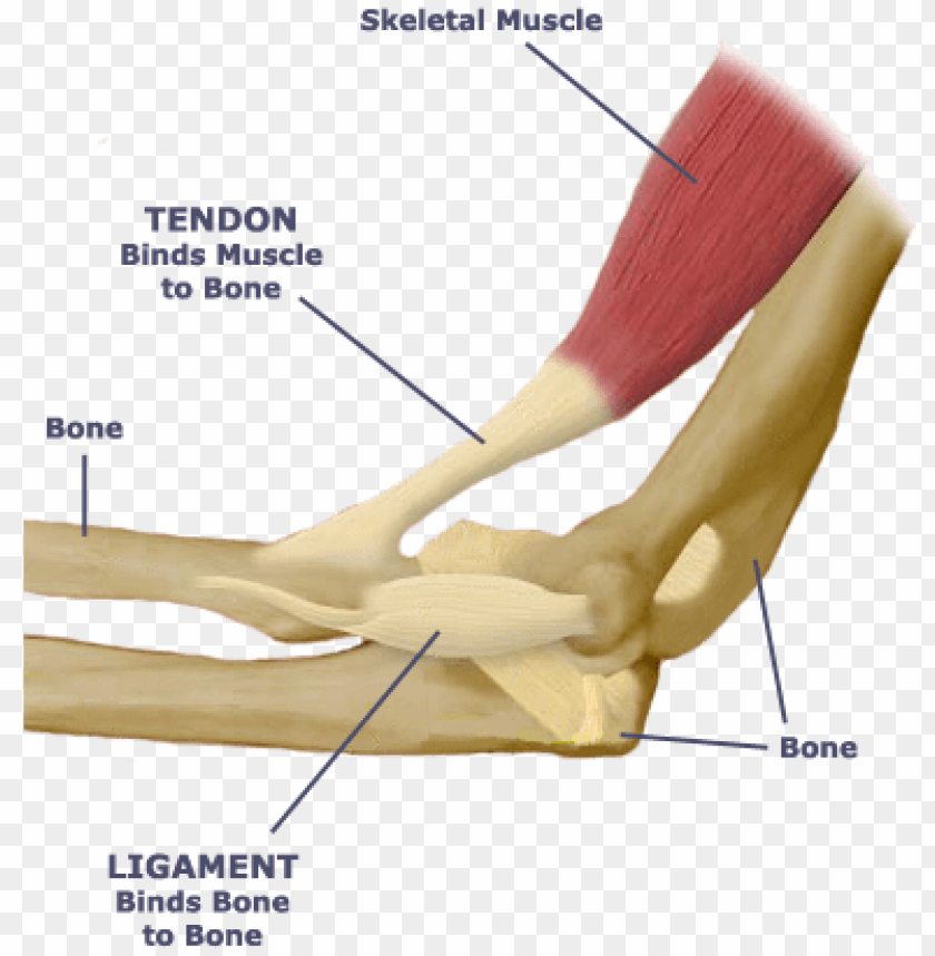
Tendon Diagram / Anatomy Of The Achilles Posterior Heel View And Ankle View. White tiger painting acrylic 97. Its muscle belly is in the forearm and then travels along the inside of the forearm and crosses the wrist. Brings trunk forward, and aids expiration. Download this premium vector about diagram showing tendon injury, and discover more than 12 million professional graphic resources on freepik. Tendons join muscles to bones.
Brings trunk forward, and aids expiration. Foot tendons and ligaments diagram tendon diagram. Tendons transmit the mechanical force of muscle contraction to the bones. The anterior cruciate ligament prevents the femur from sliding backward on the tibia (or the tibia sliding forward on the femur). Anatomy diagrams of shoulder, arm, elbow, forearm, wrist and hand.
Anatomy Of Knee from ix-cdn.b2e5.com Tendons are similar to ligaments; A tendon or sinew is a tough band of fibrous connective tissue that connects muscle to bone and is capable of withstanding tension. Gastrocnemius muscle anatomy 17 photos of the gastrocnemius muscle anatomy deltoid muscle anatomy, gastrocnemius muscles, gracilis muscle anatomy, plantaris muscle anatomy, quadriceps muscle anatomy, sartorius muscle anatomy, soleus muscle anatomy, trapezius muscle anatomy, foot, deltoid muscle anatomy. These tendon sheaths allow the tendons to glide smoothly as the diagnosis of wrist tendonitis is made by looking for the characteristic signs of this condition. Its muscle belly is in the forearm and then travels along the inside of the forearm and crosses the wrist. Possibly the most important tendon in terms of mobility is the achilles tendon. Movement at the hip joint occurs when you bend. Posted on april 3, 2019april 3, 2019.
Tendon diagrams and design force vectors.
Learn about the anatomy and physiology of tendons. The cells change shape and have more cytoplasmic organelles for increased protein production (proteogycans and collagen). Anatomy of human foot with labels on white background human anatomy for the artist: The achilles tendon is the strongest and largest tendon in the body. Posted on april 3, 2019april 3, 2019. Ankle and foot structure and actions. Tendon diagram of calf and knee. Human hand tendon diagram (page 1) hand tendons diagram muscle blank drawing these pictures of this page are about:human hand tendon diagram. The muscle belly then crosses the entire upper arm and separates into two tendons. On the anterior side of the shoulder the coracobrachialis. 1 tendons join muscles to their corresponding bones. Knee tendon diagram manual e books. Tendon, tissue that attaches a muscle to other body parts, usually bones.
The achilles tendon is the strongest and largest tendon in the body. Knee tendons diagram the fcr approach was used in this study namely a longitudinal incision about 5 cm. The foot diagram has a complex structure made up of bones, ligaments, muscles, and tendons.understanding the structure of the foot is best done by looking at a foot. White tiger painting acrylic 97. Tendon diagram simple / 8.4c:
What Is The Relationship Between The Skeletal System Tendon And Ligament Diagram Png Image With Transparent Background Toppng from toppng.com Knee tendon diagram manual e books. To bend the elbow and to turn the palm of the hand towards the sky. The two peroneal tendons in the foot run side by side behind the outer ankle bone. Tendon diagram simple / 8.4c: For more anatomy anatomynote.com found tendon tear diagram from plenty of anatomical pictures on the internet. Extensor tendon diagram 2 47 peak torque of flexor and extensor leg muscles during isokinetic. These tendon sheaths allow the tendons to glide smoothly as the diagnosis of wrist tendonitis is made by looking for the characteristic signs of this condition. Tendon diagrams and design force vectors.
Tendons that make this possible include:
The muscle belly then crosses the entire upper arm and separates into two tendons. Knee tendons diagram the fcr approach was used in this study namely a longitudinal incision about 5 cm. Allows the foot to be turned inward and also supports the arch of the foot. It is a band of fibrous connective tissues. Extensor tendon diagram 2 47 peak torque of flexor and extensor leg muscles during isokinetic. The achilles tendon is also called the calcaneal tendon. For more anatomy anatomynote.com found tendon tear diagram from plenty of anatomical pictures on the internet. 19 photos of the knee tendon anatomy diagram and name chart. Anatomy diagrams of shoulder, arm, elbow, forearm, wrist and hand. The anterior cruciate ligament prevents the femur from sliding backward on the tibia (or the tibia sliding forward on the femur). It is also capable of withstanding tension. The shoulder girdle includes three bonesthe scapula clavicle and humerus. This is as a result of compressive or tensile overload.
Tendons are fibrous cords, similar to a rope, and are made of collagen. Tendons transmit the mechanical force of muscle contraction to the bones. The achilles tendon is also called the calcaneal tendon. Tendon diagrams and design force vectors. For more anatomy anatomynote.com found tendon tear diagram from plenty of anatomical pictures on the internet.
Achilles Tendon Function Location Thompson Test Kenhub from thumbor.kenhub.com A tendon or sinew is a tough band of fibrous connective tissue that connects muscle to bone and is capable of withstanding tension. Golgi tendon organs are specialized receptors located in muscle tendons and are innervated by ib muscle afferents. It is also capable of withstanding tension. Ligaments join the knee bones and provide stability to the knee: Anatomy of human foot with labels on white background human anatomy for the artist: Without tendons, your muscles wouldn't be able to make your bones move. Foot tendons and ligaments diagram tendon diagram. The shoulder girdle includes three bonesthe scapula clavicle and humerus.
The shoulder girdle includes three bonesthe scapula clavicle and humerus.
Tendons generally have a very complex structure; Both are made of collagen. A tendon, also known as a sinew, is a fibrous tissue that helps to facilitate this movement. Learn about the anatomy and physiology of tendons. Tendon diagram simple / 8.4c: This important tendon in the back of the calf and ankle connects the plantaris, gastrocnemius, and soleus muscles to. Tendon diagram simple / 8.4c: Tendons are sometimes confused with ligaments. Tendon diagram simple / 8.4c: Brings trunk forward, and aids expiration. One tendons inserts onto the forearm bone, the radius, and the second spreads out to join the fascia along the upper part of the forearm. Posted on april 3, 2019april 3, 2019. Tendon diagram simple / 8.4c:
Share :
Post a Comment
for "Tendon Diagram / Anatomy Of The Achilles Posterior Heel View And Ankle View"


Post a Comment for "Tendon Diagram / Anatomy Of The Achilles Posterior Heel View And Ankle View"